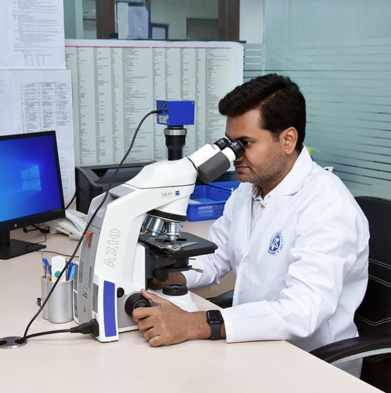
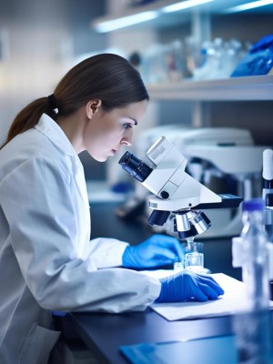
Pathology is the study and diagnosis of disease through examination of oragans, tissues,budily fluids, and whole bodies (autopsies).
study of disease
Pathology is the medical speciality that focuses on the study of diseases.
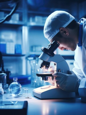
It is a broad and complex scientific field which seeks to understand the mechanisms of injury to cells and tissues, as well as the body’s means of responding to and repairing injury. Areas of study include cellular adaptation to injury, necrosis, inflammation, wound healing and neoplasia. It forms the foundation of pathology, the application of this knowledge to diagnose diseases in humans and animals.
Contact Us
Surgical Pathology

Cytopathology

Clinical Pathology

Microbiology

Screening Programme
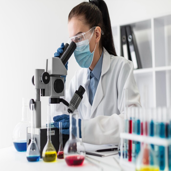
All tissues that are removed during surgery, tissues biopsies, cytological specimens & frozen sections are prepared, processed, and analyzed within our surgical pathology section. The surgical pathology section is directed and staffed by pathologists, who are specialized physicians trained to interpret microscopic tissue slides and make diagnoses. Arguably the most important of these is the diagnosis of cancer. This includes the determination of the presence or absence of cancer in patient specimens, and the determination of the severity and extent of cancer, known respectively as grade and stage.
We are equipped with a modern grossing workstation that provides a multi featured ergonomic, safe and practical work area for grossing of samples. It is the area where patient tissues taken from surgery or other procedures are received, described, and sectioned in preparation for microscopic examination. Pathologists, pathology assistants, or technicians perform the initial evaluation and processing while using appropriate physical safeguards and personal protective equipment (PPE). Most tissues received in the gross laboratory are sectioned into small pieces and placed in a formalin solution for fixation.
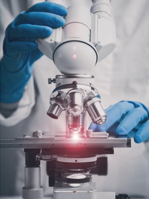
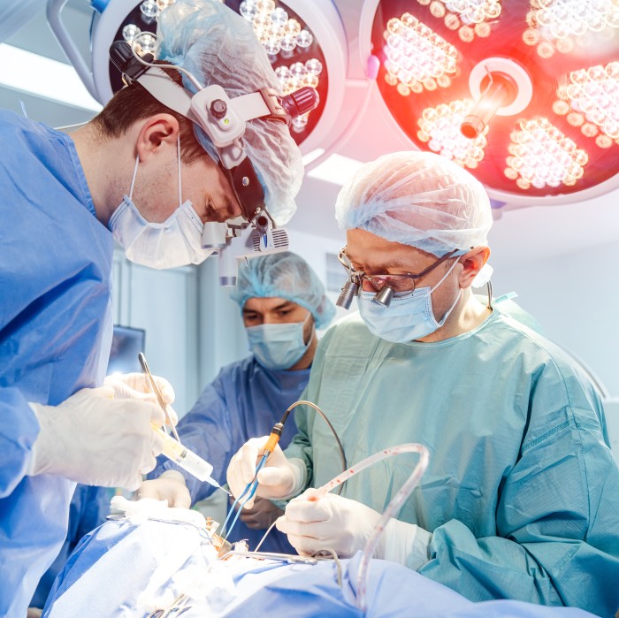
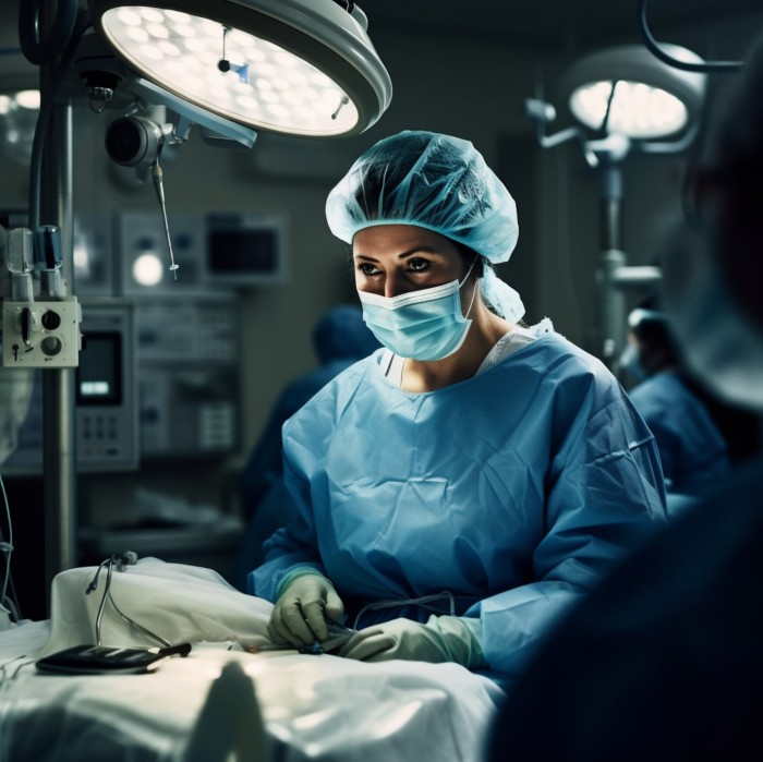
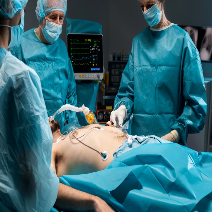
We have advanced semi-automated cryostat machine (Leica CM1520, Germany) for frozen section. It is the area where fresh tissue taken during surgery is submitted by the surgeon for immediate diagnosis by the pathologist. This is only done when the surgeon has a need for an immediate diagnosis, typically in regard to cancer. The fresh tissue is rapidly frozen and then sectioned into very thin slices using an instrument called a micro tome. The tissue slices are then placed on glass slides, stained, and examined by a pathologist using a microscope. The pathologist must provide the diagnosis to the surgeon within 15 minutes! Often this means the pathologist must make an instant decision on whether or not cancer is present in the specimen. The pathologist then reports the diagnosis to the surgeon in the operating room. The surgeon will decide how to proceed based on the immediate frozen section diagnosis.
We are equipped with fully automated microtome (Leica HistoCore AutoCut, Germany), three semiautomated microtomes (Leica, Germany), automated tissue processers & well-trained histotechnologist. It is the area where routine slides are made from the fixed tissue received from the gross pathology laboratory. The slides are stained with routine H&E stain & other histochemical special stains. These slides are presented to the pathologist for microscopic examination, diagnosis, and reporting.
Immunohistochemistry (IHC) is a widely used ancillary testing method in anatomic surgical pathology for cell classification and diagnosis and utilizes antibodies targeted against certain antigens in specific tissues and cells to facilitate determination of cell type and organ of origin which further aids in definite diagnosis of cancer.
It is the area where cytotechnologists prepare and examine tissues and body fluids for the presence of abnormal cells such as cancer cells. Then the pathologist evaluates the specimen and provides a final diagnosis. It
is the area where cytotechnologists prepare and examine tissues and body fluids for the presence of abnormal cells such as cancer cells. Then the pathologist evaluates the specimen and provides a final diagnosis.
It is the area where cytotechnologists prepare and examine tissues and body fluids for the presence of abnormal cells such as cancer cells. Then the pathologist evaluates the specimen and provides a final diagnosis. It is the area where cytotechnologists prepare and examine tissues and body fluids for the presence of abnormal cells such as cancer cells. Then the pathologist evaluates the specimen and provides a final diagnosis.
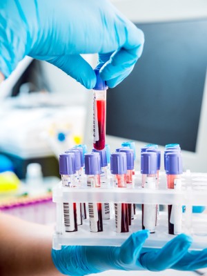
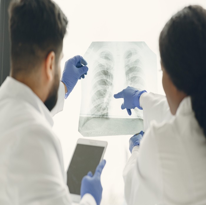
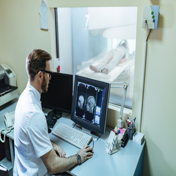
Clinical pathology (Laboratory medicine) is a medical specialty that is concerned with the diagnosis of disease based on the laboratory analysis of bodily fluids such as blood and urine, and tissues using the tools of chemistry, microbiology & hematology. Clinical pathologists work in close collaboration with medical technologists, hospital administrations & referring physicians to ensure the accuracy and optimal utilization of laboratory testing.
We are equipped with fully automated hematology analyzer (Horiba Yumizen H550, Japan) & semi automated coagulation analyser (Tcoag KC1 Delta, Ireland). It is the area where blood samples are analyzed for quantitative & qualitative abnormalities of blood cells & plasma. Then the pathologist evaluates the specimen and provides a final diagnosis.

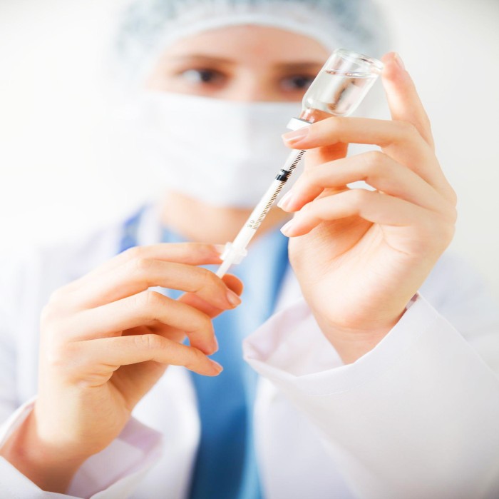
We are equipped with fully automated biochemistry analyzer (Siemens Dimension EXL 200, Germany), compact automated immunoassay system (Biomerieux Mini Vidas) based on the Enzyme Linked Fluorescent Assay (ELFA) principle & electrolyte analyzer (Easylyte Expand). It is the area where blood serum/plasma samples are analyzed for quantitative & qualitative abnormalities.
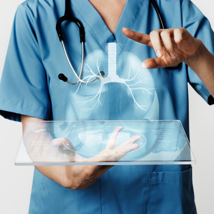
We operate a state-of-the-art automated microbiology analyser, specialising in the examination of diverse tissue, body fluids, and blood samples. Our advanced facility is dedicated to the precise detection of pathogenic microbes and assessing their sensitivity to antibiotics. Through cutting-edge technology, we ensure accurate and efficient microbiological analyses, contributing to informed diagnostic decisions and tailored treatment strategies.
Screening is a way of finding out if people have a higher chance of having a health problem, so that early treatment can be offered or information given to help them make informed decisions.
What is screening?
Screening is a way of identifying apparently healthy people who may have an increased risk of a particular condition. The Rajkot Cancer Society offers a range of screening tests to different sections of the population. The aim is to offer screening to the people who are most likely to benefit from it.
What types of screening are offered by Rajkot Cancer Society?
Currently we provide screening programs for cervical & breast cancer in collaboration with Kundariya Cancer Prevention Foundation (KCPF).
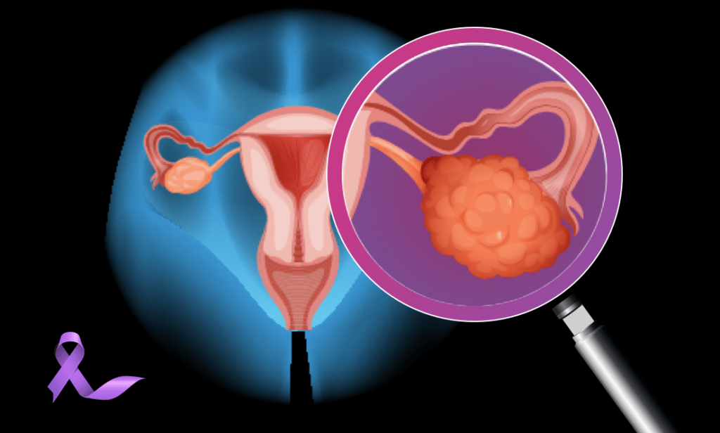
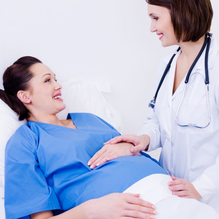
Cervical screening is offered to all women with a cervix aged 25 to 64 to check the health of cells in the cervix by conventional PAP smears. It is recommended every 3 years for those aged 25 to 49, and every 5 years from the ages of 50 to 64.
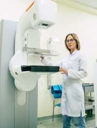
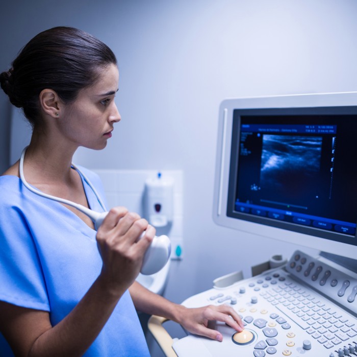
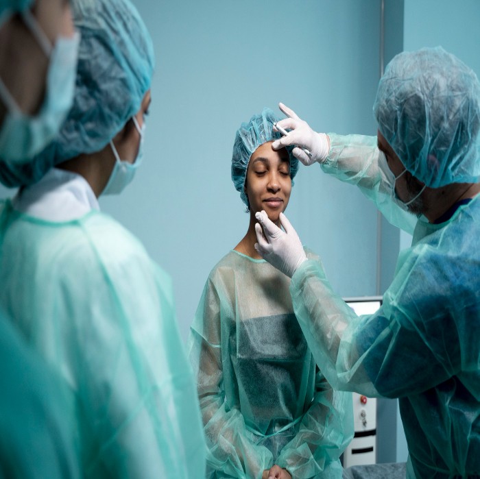
Breast screening is offered to women aged 30 to 70 to detect early signs of breast cancer by clinical examination by surgeons, sonomammography & cytopathological & histopathological examination in relevant cases.

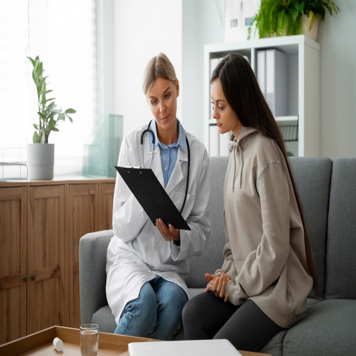
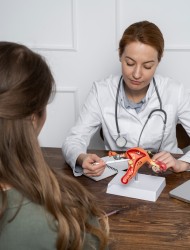
Schedule your doctor's appointment at Rajkot Cancer Society for expert and compassionate care, ensuring personalized attention to your health needs.